The MCP has adopted various tracer injection strategies in order to compile the most accurate and reliable connectivity data. Data are systematically collected in 8-week old adult male C57Bl/6 mice. Following tracer injections, the brain is sectioned along the coronal plane and immunohistologically processed for tracer visualization. Each section is 50 µm thick and one out of every four sections across the entire brain is presented in a single series in the iConnectome viewer. All sections are counterstained with the fluorescent Nissl stain NeuroTrace Blue to provide cytoarchitectural details. The fluorescent Nissl is converted to brightfield Nissl in the image processing phase.
(1) Double coinjections. In this strategy, two injections are made into two separate delineated regions of a mouse brain. Each injection contains a mixture of an anterograde and a retrograde tracer. Phaseolus vulgaris leucoagglutinin (PHAL; anterograde) and cholera toxin subunit b (CTb; retrograde) are coinjected, while biotinylated dextran amine (BDA; anterograde) is coinjected with Fluorogold (FG; retrograde).
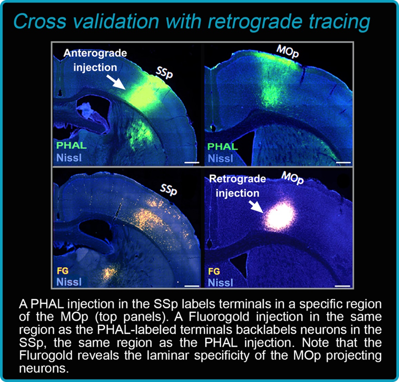
Importantly, retrograde tracers provide relevant information regarding the input of structures, but they are also used to validate the anterograde labeling results.
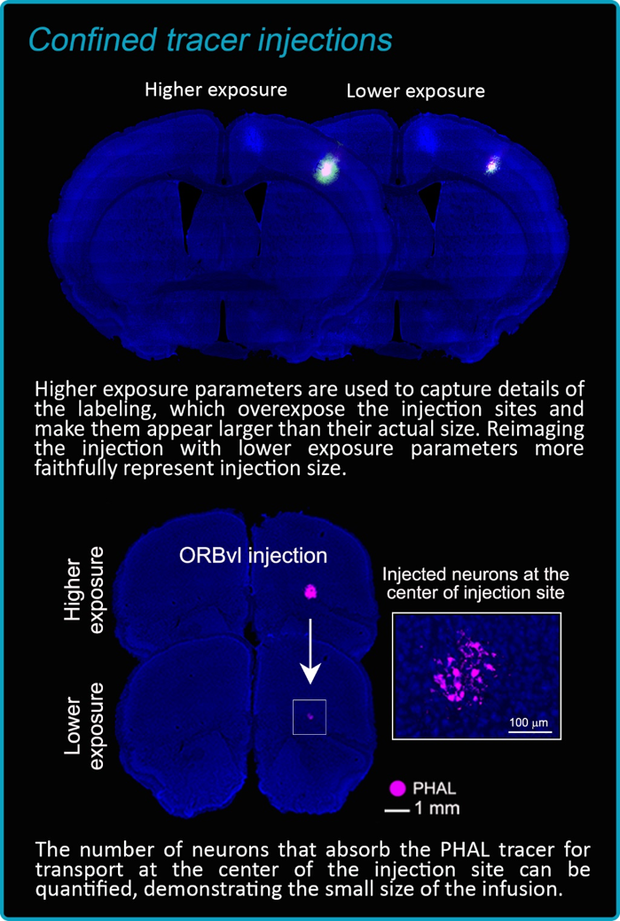
Small tracer injections confined to a single anatomically delineated region also play an important role in acquiring the most reliable connectivity data.
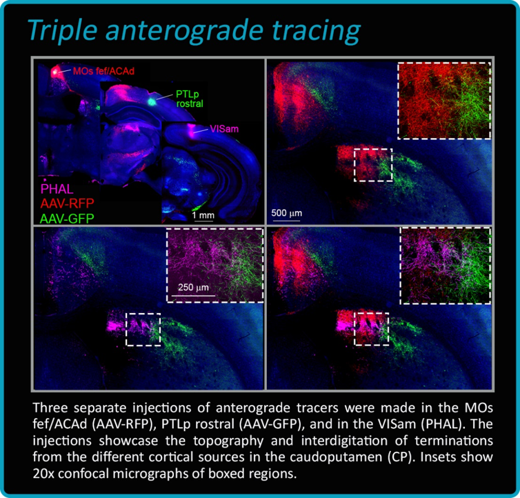
(2) Triple anterograde tracing. In this strategy, three different anterograde tracers are injected into various regions of interest to highlight the spatial correlation of topographic projections from different neural structures. Typically, PHAL and adeno-associated viruses (AAVs) that express either green (AAV-GFP) or red (AAV-RFP) fluorescent protein are injected in a single animal.
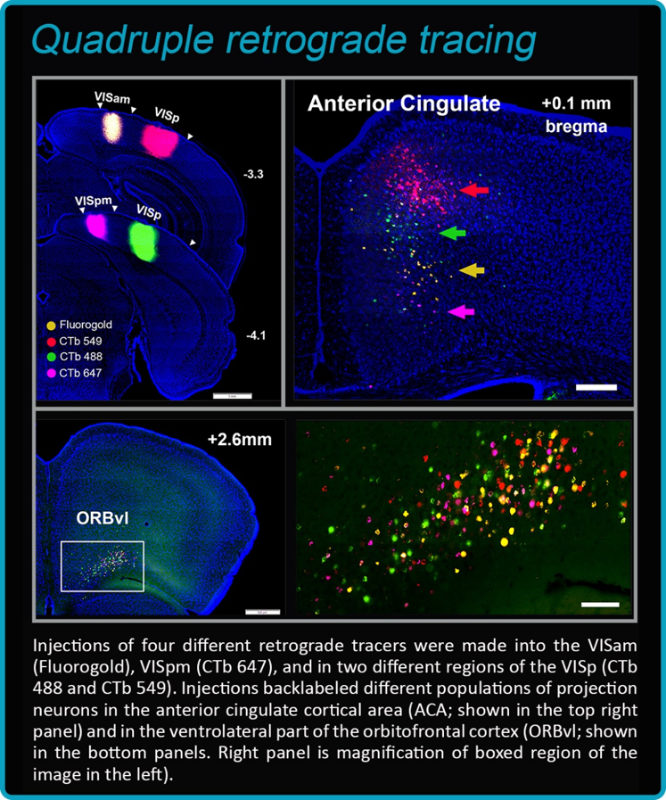
(3) Quadruple retrograde tracing. Individual groups of cells located within a single delineated structure have differentprojection targets. To reveal these different populations of neurons with unique projection targets and their spatial relationship to one another, four different retrograde tracers are injected into the targets of different projection neurons. The retrograde tracers include a combination of FG, CTb conjugated with AlexaFluor 488, 647, or 549, and rabies that express green or red fluorescent protein.